

- Home
- Our Work
- Normal Subjects
- Talks
- Interviews
- FAQs
- Collaborators
- CCSVI Protocols

- History
- Contact Us
|
Collaborate with MS-MRI
Since the summer of 2010, we have processed more than 2,000 MS cases. We are constantly data mining these images and looking for important physiological data on MS and CCSVI. We invite you to send any relevant information such as research papers on MS research that you think may be of help related to the CCSVI theory. Please also read the history section on this web page.
As paraphrased by Dr. George B. Hassin of Chicago in 1935, when reviewing the more risky treatment of MS with sympathectomies: "...multiple sclerosis is such a distressing, hopeless condition that any therapeutic measure that appears promising should be given careful consideration, regardless of the difference of opinion as to the pathophysiologic features of this disease" [Wetherall]. Wetherell F. Cervicodorsal sympathectomy in multiple sclerosis. Arch Neurol Psych 1935; 34: 99-110. |
|
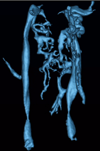
The CCSVI (Haacke) Protocol With the current interest in chronic cerebral spinal venous insufficiency (CCSVI) and the role of abnormal venous flow and its effects on brain perfusion and brain function, Prof. Haacke has designed a full protocol for studying the brain, neck and spine anatomy and function. This CCSVI protocol contains the usual conventional neurological protocol to study anatomy, measure lesion load and iron load, measure the presence of chronic and acute lesions, (in some cases perfusion weighted imaging for cerebral blood volume, flow and mean transit time), as well as the vascular components of MR angiography, MR venography, quantitative flow imaging, and more recently the potential to study oxygen saturation. This comprehensive protocol is critical for the patients because it provides a baseline from which to follow the patient longitudinally post treatment. WHY IMAGING IS CRUCIAL FOR MS PATIENTS PRE AND POST TREATMENT There has been a great deal of discussion on the role of imaging pre and post treatment of CCSVI. It is important to understand that every person is different and the geometry of the venous system is different as well. Bearing in mind that MRI provides complete 3D information of the vascular system and the anatomy of the body, it has been suggested that MRI is critical for the following reasons: 1) It provides key neurological information for the patient and the neurologist following the patient such as the presence or absence of lesions which are markers for the state of the disease 2) It provides a means to monitor iron content. 3) It can visualize arteries and veins in the head, neck, spine and aortic arch which are key elements in treatment planning for the interventionalists. 4) It provides a means to measure blood flow in each major vessel everywhere in the body. 5) It can quantify quantitative information on perfusion to the brain tissue. 6) These data serve as a control prior to treatment. The information is objective and quantifiable. This is especially critical in case the patient suffers from restenosis. These flow measurements can be repeated as can the MR angiograms non-invasively. MRI itself is a non-invasive safe imaging procedure. 7) It can be used for follow up whether or not there is restenosis. Longitudinal follow up could well serve as an indicator as to the status of the disease, i.e., the presence or absence of new lesions, a stable vascular system, changes in iron content, etc. 8) These records become a key component in comparing to the venous angioplasty and patient outcomes. |