

- Home
- Our Work
- Normal Subjects
- Talks
- Interviews
- FAQs
- Collaborators
- CCSVI Protocols

- History
- Contact Us
|
Collaborate with MS-MRI
Since the summer of 2010, we have processed more than 2,000 MS cases. We are constantly data mining these images and looking for important physiological data on MS and CCSVI. We invite you to send any relevant information such as research papers on MS research that you think may be of help related to the CCSVI theory. Please also read the history section on this web page.
As paraphrased by Dr. George B. Hassin of Chicago in 1935, when reviewing the more risky treatment of MS with sympathectomies: "...multiple sclerosis is such a distressing, hopeless condition that any therapeutic measure that appears promising should be given careful consideration, regardless of the difference of opinion as to the pathophysiologic features of this disease" [Wetherall]. Wetherell F. Cervicodorsal sympathectomy in multiple sclerosis. Arch Neurol Psych 1935; 34: 99-110. |
|
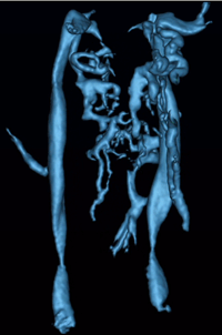
Chronic Cerebral Spinal Venous Insufficiency (CCSVI). Ultrasound (US) can image the vessel and flow in real time. As such, it can monitor the motion or lack of motion of a valve throughout the cardiac cycle. However, US does not have the full scope of 3D coverage that MR does nor the in-plane resolution although through plane resolution can be quite high for the vessel wall. Contact your neurologist and discuss the options with them. Also, look for the Institute closest to you that has IRB or REB approvals to collect such data. You should ask the site where you are scanned if they will provide you with a CD to take to your neurologist and radiologist. The MRI scan usually lasts one to one and a half hours. Many normal people have only one jugular vein. We do not know. We do not know. The higher field magnets would produce a higher torque on magnetic objects. Torque can cause an object such as an implanted stent or clip or anything else like this to rotate (move) inside the body which could damage the surrounding tissue. More worrisome would be the different radiofrequency interactions that would take place in the presence of a metallic object when running certain MR scans. This could cause local heating and again cause damage to the surrounding tissue. Therefore, a given stent should be tested at 1.5T or 3T if you will be scanned on one of these systems. The safety book for this is updated every year. It is my guess that 3T will remain the state-of-the-art clinical system for at least 20 years. These human imaging systems are expensive, a 3T system costs about $3M and a 7T system costs about $7M. Since 7T systems are very high field, the problems mentioned at the beginning of this paragraph are potentially worse. And given their great expense, and the fact that they are still under development, 7T is unlikely to become a standard system anytime soon, although they are great research tools. We do not know if iron chelators help or not. An MRI would have no effect on iron in the brain that we know. We do not know the answer to this question at this time. We do not know the answer to that question at this time. The current CCSVI MRI protocol that Dr. Zivadinov reported includes both MRV and flow quantification. MR flow quantification is as good as and perhaps even better than ultrasound. With these two features together, MR can catch a lot of the abnormal vessels. Further, MRI can create full 3D vascular information from the aortic arch to the top of the brain. Doppler is also more operator dependent than the MRI. But ultrasound can image the valves and septum in the veins which MRI can not do. So together they make a good combination with flow acting as a common link between them. In summary, both ultrasound and MRI are very important. As in any technological applications, imaging methods will only get better over time and our ability to diagnose CCSVI will get better. The imaging methods only suggest CCSVI. MRI is a critical assessment tool, especially from the neurological perspective. MRI can measure atrophy, iron content and if the veins are patent. This remains to be determined. Yes and no. Arteries and veins can be imaged without contrast agent. But the ability to image the vessels is improved by using a contrast agent. Also, flow quantification can be done without a contrast agent and measuring iron can be done without a contrast agent. |